Hip Preservation Surgery
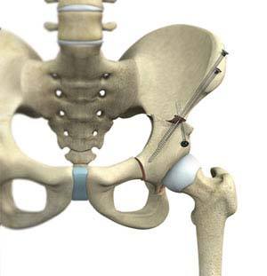
The hip joint is one of the body's largest weight-bearing joints and is the point where the thigh bone (femur) and the pelvis (acetabulum) join. It is a ball and socket joint in which the head of the femur is the ball and the pelvic acetabulum forms the socket. The joint surface is covered by a smooth articular cartilage that cushions and enables smooth movements of the joint.
Hip pain has become a common problem particularly in sportspersons involved in vigorous activities. The main reason behind the development of hip pain is due to changes in the shape or structure of the ball and socket type of hip joint.
The treatment options for the management of hip disorders such as excruciating hip pain and hip dysfunction, in young patients, have been limited. However, the newer minimally invasive techniques have been found to be beneficial in these patients and these also avoid the need of hip replacement.
Hip arthroscopy is an excellent surgery that has helped many patients restore their hip function and alleviate pain originating from their hip. Hip arthroscopy, is a minimally invasive surgery that is performed through very small incisions to evaluate and treat a variety of painful hip conditions. An arthroscope is a pencil-sized instrument that has a small lens and lighting system at one end. The arthroscope magnifies and illuminates the structures inside the body with the light that is transmitted through fibre optics. It is attached to a television camera and the internal structures are seen on the television monitor.
A variety of painful conditions can be treated using this technique. Many patients are able to return to activities that they were unable to do for years because of pain and limited range of motion.
Hip arthroscopy may be indicated in the following conditions:
- Debridement of loose bodies: Bone chips or torn cartilage debris cause hip pain and decreased range of motion and can be removed with hip arthroscopy.
- Repair of torn labrum: The labrum lines the outer edge of the “socket” or acetabulum to ensure a good fit. Tears can occur in the labrum causing hip pain.
- Removal of bone spurs: Extra bone growth caused by injury or arthritis that damages the ends of the bones cause pain and limited joint mobility.
- Restoration and reconstruction of joint surfaces: Injury to the articular cartilage can lead to arthritis. If treated early by arthroscopy, the development of arthritis may be delayed or even prevented altogether.
- Evaluation and diagnosis: Patients with unexplained pain, swelling, stiffness and instability in the hip who had no success with non-operative treatments, may undergo hip arthroscopy for evaluation and diagnosis of their condition.
Hip preservation surgeries are also indicated in patients with cartilage defects, to preserve the hip and to restore its functionality. The articular surfaces of the hip joint are lined by a cartilage, known as articular cartilage. It has a smooth surface which allows the articular surfaces to slide over one another with minimal friction. Articular cartilage is often damaged by injury or normal wear and tear. Articular cartilage, when damaged or worn away, the affected joint becomes painful, stiff, and has limited range of motion. As the articular cartilage has limited ability to heal by itself, surgical repair may be required to stimulate the growth of new cartilage. Articular cartilage restoration relieves pain, improves function and can delay or prevent the onset of arthritis in the joint. Cartilage restoration can be achieved using different techniques.
- Microfracture surgery: Microfracture surgery is appropriate for patients having single lesion and healthy subchondral bone. Microfracture can be done using an arthroscope- a long, thin device with a tiny camera attached at the end to see inside your hip. Your surgeon uses a small pointed tool called an awl to make very small holes, microfractures, in the bone underlying the cartilage called subchondral bone. This stimulates the healing process by increasing the blood flow to the surface which brings in new cells that build new cartilage.
- Drilling: This technique involves use of a surgical drill or a fine wire to make multiple small holes through the damaged area to penetrate the subchondral bone. This generates a healing response within the defect. As the heat produced during drilling can cause injury to some of the tissues, it is considered less accurate than microfracture.
- Abrasion arthroplasty: Abrasion arthroplasty can be done using an arthroscope. This technique is similar to drilling, rather than drills or wires, high speed burrs are used to remove the damaged cartilage and to penetrate the subchondral bone.
- Autologous chondrocyte implantation: Autologous chondrocyte implantation (ACI) is a two-step procedure. The first step is an arthroscopic surgery to remove the tissue containing healthy cartilage cells from an area of the bone that does not carry weight. These cells are cultured and multiplied in a laboratory over a 3- to 5- week period. In the second step, the newly grown cells are implanted through an open surgical procedure, or arthrotomy. During this surgery, a periosteum, a layer of bone-lining tissue is sewn over the area of damaged cartilage. Once the area is sealed with fibrin glue, the cultured cartilage cells are injected underneath the periosteal cover.
This procedure is appropriate for patients with a single lesion of large area. As the patient’s own cells are used, chances of rejection is not a concern. However, it is a two-step procedure, requiring large incision and lengthy recovery.
- Osteochondral autograft transplantation: In this procedure, healthy cartilage (graft) taken from a non-weight-bearing area is transferred to a damaged area of the hip. The graft is taken as a cylindrical plug of cartilage and underlying bone. Then the graft is matched with the damaged area and transplanted into place. This leaves a smooth cartilage surface in the joint. Transplantation may be performed using a single plug or by mosaicplasty where multiple plugs are used.
This technique is used for patients with small areas of cartilage damage because of the limited availability of the healthy cartilage from the same joint.
- Osteochondral allograft transplantation: During this technique, tissue graft taken from a cadaver donor, known as allograft is used to repair the damaged cartilage. Allograft may be used if the cartilage defect is too large for an autograft.
Rehabilitation
Mr. Carton may recommend physical therapy following any of the hip preservation surgeries to strengthen the joint and the muscles and help restore mobility to the hip joint.