The Importance of Comprehensive Cam Correction
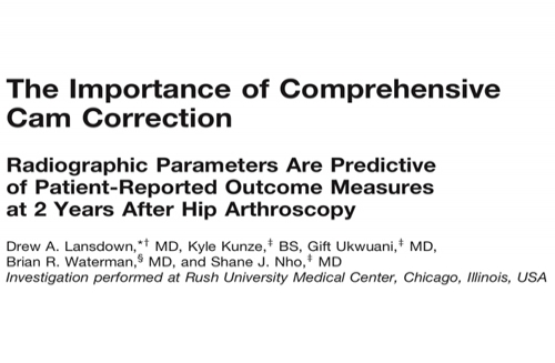
The American Journal of Sports Medicine
(2018) 46(9): 2072-2078
Critical Appraisal:
This Journal Club meeting critically appraised the above paper which focuses on the importance of optimal and comprehensive cam resection. Adequate resection of bony-contributing pathology is a vital component of hip arthroscopy, and the authors do highlight in the introduction that residual bony deformity presence is one of the main reasons for poorer outcomes, contribute to the development of OA and decreased survivorship in young adults. Residual deformity is also the most common reason for undertaking revision hip arthroscopy surgery. From this point of view, the study does address a clearly focused question
Recruitment Process/Patient Selection:
This is a retrospective analysis of prospectively collected data from a consecutive series of patients undergoing hip arthroscopy for femoroacetabular impingement (FAI) by one single high-volume surgeon at one institution. The selection process was considered to be appropriate – the diagnosis of FAI was made using a triad of findings (patient history, physical examination and imaging findings), inclusion/exclusion criteria applied were consistent with currently available indicators and contra-indicators for hip arthroscopy for FAI. The authors of this paper however did include Tonnis Grade up to and including Grade 2 – a grade which can indicate the presence of quite moderate narrowing of the joint space. In much of the hip arthroscopy outcome-based literature Tonnis Grade 2 would also be an exclusion criteria. The presence of these cases may therefore be influencing the results as typically poorer pre- and post-operative outcome scores would present in these patients. The authors also have not provided any breakdown of these patients in terms of the different Tonnis Grades present among the cohort overall.
The cohort in this study consisted predominately of females (64.4%) which is in direct contrast to the gender mix of patients we would see at our clinic with true FAI (predominantly young male patients). In our experience, the vast majority of females do not have cam deformities and are symptomatic mainly as a consequence of mild dysplasia, instability or pincer impingement, only 70% of our males have evidence of cam impingement. However, no detail was provided in this study to the radiologic angles of males and females separately and the degree of correction with respect to gender. Due to the significant differences in outcome, presentation and pathology, between male and females, more detail should have been provided.
Radiographic Analysis
Questions were raised as to whether the radiographic measurements truly reflect the presence of a pathologic cam deformity. In particular the description by the authors of a cut-off value of 50 degrees to represent a cam deformity on any x-ray view. It has previously been reported that an alpha angle >65 degrees on the AP view and >55 degrees on the Dunn view would represent the presence of a measurable cam deformity. In our clinic these are the x-ray parameters we utilise to define abnormal femoral head neck offset.
The measurement of 50 degrees on all views may significantly overestimate the percentage of people designated with a cam deformity and as such include many patients who would not be considered to have abnormal femoral pathology in other surgical centres.
Furthermore, particularly in females with Tonnis Grade 2 present, (indicating the presence of a wearing hip), osteophytes may develop which could replicate a cam deformity (by increasing the alpha angle significantly). Including Tonnis Grade >2 therefore may inadvertently increase the inclusion numbers to the study.
Considering this is an x-ray focused paper, details on the method in which x-rays were taken, standardised and measured was lacking and potentially is a major flaw in the methodology. There were also no details on whether certain people were excluded based on excessive rotation/tilt of either pre- or post-operative x-rays as this would lead to significant inaccuracy of measurements. It would appear that this was not taken into consideration, given all cases meeting the defined inclusion/exclusion criteria, apart from those lost to follow-up, were included in the study.
Additionally, there is no mention when these x-rays were taken post-operatively (the same day, next day, weeks-months later, or at the same time as the 2 years follow up). This is an important point which should have been mentioned. For instance, if an x-ray is taken the day of or after surgery for example, patients may still be sore and obtaining standardised x-rays with the correct degree of limb rotation, flexion, etc would be difficult and this could further lead to inaccurate x-ray images and measurements.
Surgical Technique
Overall, this is standardised. There are some differences in technique when we think of the surgery performed at our own clinic. For example, T-capsulotomy performed in this study compared to an inter-portal capsulotomy performed routinely at our clinic. The authors did make reference to cartilage quality as normal, mild, moderate, severe and the presence of delamination was recorded, however the impact of this grading on patient outcomes was not clearly defined in the results. This grading system was not used to evaluate outcomes or radiographic parameters in the results section, therefore unsure as to why it was mentioned in this section.
Rehabilitation
It was considered that the rehabilitation post-operatively was quite conservative, particularly when we would compare to the protocol in use at our clinic. The authors recommend restricted weightbearing on the operated limb for 3 weeks post-operatively. However appropriate removal a cam deformity is unlikely to interfere with the natural strength of the femoral neck and we advise full weight-bearing as comfortable straight after surgery with removal of crutches altogether after 5 days. A large proportion of bilateral cases coming through our clinic will have their surgery 1 week apart with no issues mobilising post-operatively after either surgery. We consider that restriction of joint and muscle movement to comply with a more conservative post-operative regime may result in the patient finding it harder to regain these movements at a later stage and may increase the risk of developing post-operative adhesions.
Results
78-84% follow-up rate at 2 years reported is very acceptable. Not all patients have completed all of the outcomes, and it would have been helpful if the description or breakdown of the outcomes and radiographic parameters specific to the different demographics within the cohort had been included. In addition, the authors do not present any baseline data for patients who were followed up compared to those with no outcomes, to demonstrate to the reader that these groups were not grossly different.
Figure1 –while visually we can see there is an obvious and statistical difference in the pre-op to post-op scores, it would be more impactful if the actual mean values with SD was also supplied in an accompanying table, or at least within the text in the results section, however this was not supplied.
In this study there was a mean resection from 78 degrees pre-op to 44 degrees post-op which is almost a 50% reduction in their alpha angle (and similar on all other x-ray views). This mean cam resection difference is very considerable. In our practice, our mean AP alpha angle improves from a mean of 64degrees pre-op to 44degrees post op (we consider an angle >65 degrees as indicative of abnormal morphology on this AP view). We have a similar reduction in mean alpha angle of the Dunn view (59To 46With >55 degrees considered abnormal).
The 2 year post-operative mHHS of 79.1 is lower than in our series of patients were we have reported mHHS of 92at a similar timepoint from surgery despite having a similar average age of our patients.
The outcome of surgery depends on good patient selection and appropriate correction of both pincer and cam deformity and optimal management of the labrum and capsule. At our clinic we perform chondrolabral preserving labral repair and utilise a more conservative inter-portal capsulotomy and repair. Our primary focus is on the anatomical restoration of the acetabular rim and chondrolabral junction and in particular removing anterior pincer deformities. This focus on the anatomical restoration of the acetabular component of FAI we believe leads to the optimal post-surgical outcomes observed and reported at our clinic.
The overall aim of this study was to try and correlate the extent of cam deformity with the post-operative outcomes and the extent of the resection as to whether you will do well or poorly after surgery. The authors detail the specifics of their correlations with the use of rho values accompanied with p-value level of significance. These p-values ultimately signal how significant the measurement of the correlation is. In Table 1 the highest correlation reported is with the HOS_SSS for pre-operative alpha angle (Rho -0.24) which is extremely weak when trying to substantiate the overall message in this study. Where there are high significant values, this simply indicates the Rho calculation is accurate. However, this does not substantiate association. The presence of significant correlation does not imply causation. So, if you have a high pre-op alpha angle and a poor post-op outcome, this does not mean that the high pre-op alpha angle causes the low score. There could be a number of other factors including additional intra-operative procedures (in particular labral repair) or findings which may be more strongly implicated in the outcome. One of the issues with the results and discussion of this paper is that an inaccurate message is being delivered i.e. that the observed correlations in alpha angles predict outcomes, which is unlikely to be the case. The strength of the significant findings reported in the study, for one, cast inconclusiveness to this message. Elaboration therefore on the meaning of the significance of the findings (i.e. the strength of the correlations) should be made clearer in the discussion.
Main Points/Summary
The general consensus was that more information about the alpha angles should have been provided specific to different groupings within the cohort (e.g.gender breakdown, age group breakdown, Tonnis grade breakdown, missing cases demographic vs followed-up cases demographics breakdown, etc).
Significant correlations should not imply causation, particularly where these correlations are poor/weak. The weakness of the correlations with respect to outcomes was not clearly highlighted.
While a cam may be important and potentially causative to patient symptoms when present, this paper appears to attempt to try and back up the generalised line of thinking that the cam is the primary pathology –which, if we compare to our own high-volume clinic, is not the case. In many cases the cam is a secondary pathology and the main source of the problem is located at the rim of the acetabulum and chondrolabral junction.
Critical Review Panel:
The Hip and Groin Clinic, UPMC Whitfield, Ireland:
Mr. Patrick Carton MD FRCS (TR&Orth) FFSEM, Consultant Hip Surgeon
Mr. David Filan, Research AssistantMs. Karen Mullins, Research Assistant
Mr. Derek O’Neill, Chartered Physiotherapist
Ms. Sarah Coughlan, Chartered Physiotherapist
Ms. Jenny Walsh, Chartered Physiotherapist
Mr. Michael Bowler, Chartered Physiotherapist